What is a brain MRI scan? Why does an MRI scanner make different noises? And what do the different brain MRI scan sounds mean?
Thank you for joining us as we examine another video in our MRI scan sounds series. In this article, we break down the brain MRI scan protocol to examine the purpose of each pulse sequence.
Brain MRI Scan Sounds Explained
Each pulse series includes a sample of the MRI scan sound accompanied with a brief description, and the images produced by each MRI scan sequence.
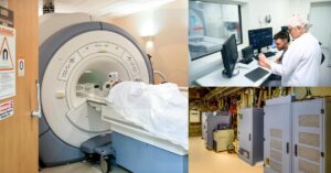
Routine Brain MRI scan performed on a GE Signa Excite 1.5T MRI scanner. Make sure to check out our MRI scan sounds playlist to view additional MRI scan sounds videos. We feature MRI scan sounds from every GE 1.5T MRI and 3T MRI scanner models. Links will be provided in the additional resources section.
Let’s get started with the first pulse sequence you hear during a brain MRI scan: the auto prescan tuning sequence.
1. APS (Auto Prescan)
MRI scan sounds begin at 00:53
Auto prescan tuning sequence performed prior to each scan series to determine optimal center frequency, RF output, and offset values. APS sequence does not produce images.
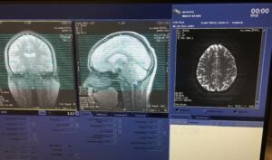
2. Three-Plane Localizer
MRI scan sounds begin at 01:22
The 3 plane localizer produces reference images in the axial, sagittal and coronal plane. MRI technologists use the images produced to plan the slice selection in the series to follow.
3. Sagittal T1 FLAIR Brain MRI
MRI scan sounds begin at 01:59
Sagittal T1 FLAIR MRI series is a fast inversion recovery sequence. It achieves better tissue contrast-to-noise ratio as well as better signal to noise in a shorter amount of time when compared to a spin echo scan sequence.
Inversion recovery imaging utilizes a spin echo pulse sequence that starts with a 180 degree inversion pulse. This creates inverse proton magnetization in the longitudinal plane. Secondly, a 90 degree pulse follows the inversion pulse.
The period of time in between the 180 degree and 90 degree pulse is known as (TI) Inversion Time. Inversion time, or TI, is the primary factor that determines image contrast.
TI is more easily defined as the amount of time it takes tissue to recover from the longitudinal plane to the transverse plane.
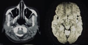
4. Axial DWI Brain MRI
MRI scan sounds begin at 02:48
Axial DWI EPI scan, commonly referred to as Diffusion weighted imaging, is a single shot echo planar imaging protocol sequence. DWI MRI scans differentiate tissue with restricted diffusion from tissue with normal diffusion coefficients based on the Brownian motion of the molecules in the tissue. Click the link on the top right to learn more about DWI MRI scans. Diffusion weighted imaging is optimized to differentiate tissue in the brain.
DWI utilizes the gradient coil to provide an echo, as opposed to relying on the RF pulse echo. Additionally, DWI MRI uses partial k-space reconstruction algorithms to reduce scan times.
5. Axial T2 FSE- XL90
MRI scan sounds begin at 03:36
The Axial T2 FSE-XL pulse sequence is an accelerated fast spin echo scan that applies optimized RF pulses to shorten echo space. This results in reduced edge blurring, enhanced tissue contrast, and faster scan times. FSE-XL scan sequences are primarily used in cardiac, abdominal and brain MRI scan sequences. Blurring correction and blood suppression techniques help to improve image quality in cardiac, kidney, brain, and other similar techniques.This scan sequence uses increased RF output as compared to the FSE sequence, expect increased MRI scan noise levels during this pulse sequence..
6. Axial T1 Spin Echo
MRI scan sounds begin at 04:26
The Spin Echo pulse series is a single plane imaging sequence which consists of a 90 degree excitation pulse with at least one 180 degree refocusing pulse.
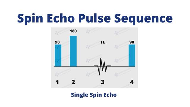
The 90 degree excitation pulse provides transverse magnetization, while the 180 degree pulse flips the protons to provide spin echo signals.
7. Axial T2 FLAIR
MRI scan sounds begin at 05:16
Axial T2 FLAIR is a fast inversion recovery technique based on the null point of cerebrospinal fluid. T2 FLAIR produces a bright CSF signal which helps to differentiate T2-weighted image structures next to fluid filled structures.
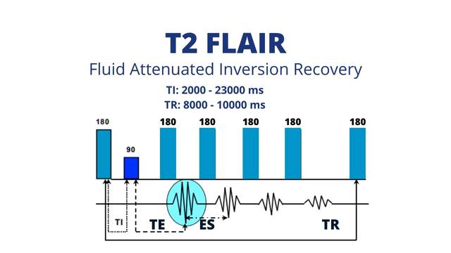
The Inversion time is the primary driver for the image contrast. The TI value should match the time it takes for CSF tissue or structure to relax, approximately 2000-2300 ms.
8. Coronal T1 FSE-XL 90
MRI scan sounds begin at 06:07
The Coronal T1 FSE-XL pulse sequence provides the same imaging capabilities equal to Axial T1 FSE-XL. The primary difference between axial and coronal being gradient pulse direction. This series provides a perpendicular view of the anatomy.
9. Coronal T2 GRE
MRI scan sounds begin at 07:11
Lastly, gradient echo pulse sequences serve as general purpose PSDs used to produce 2D and 3D reconstructions. GRE uses short TR and flip angles of less than 90 degrees to excite only a portion of the longitudinal magnetization.
Conclusion
The “Routine Brain MRI” protocol is anything but routine. Each MRI sounds and scan sequence is highly specialized to highlight the tissue and fluid in the brain.
Thank you for reading! We hope you found this to be a helpful article and video. Consider subscribing to our YouTube channel receive a notification every time we release new content.
Want to learn more? Click here to see how MRI works or make a selection from one of our helpful resources below.
Related Resources
Quick Navigation Links
Join the Medical Imaging Source Community!
Subscribe To Our Newsletter To Stay Up To Date With The Latest News, Exclusive Offers, And Giveaways!
The information provided by MRIPETCTSOURCE (“we,” “us,” or “our”) on https://www.medicalimagingsource.com (the “Site”) is for general informational purposes only. All information on the Site is provided in good faith, however we make no representation or warranty of any kind, express or implied, regarding the accuracy, adequacy, validity, reliability, availability, or completeness of any information on the Site. UNDER NO CIRCUMSTANCE SHALL WE HAVE ANY LIABILITY TO YOU FOR ANY LOSS OR DAMAGE OF ANY KIND INCURRED AS A RESULT OF THE USE OF THE SITE OR RELIANCE ON ANY INFORMATION PROVIDED ON THE SITE. YOUR USE OF THE SITE AND YOUR RELIANCE ON ANY INFORMATION ON THE SITE IS SOLELY AT YOUR OWN RISK.