
What Is MRI? Magnetic resonance imaging is a medical imaging marvel that produces a symphony of sounds to create high definition images of the anatomy without ever needing to perform invasive surgery. Thank you for joining us as we explore magnetic resonance imaging.
This article details what an MRI is and, most importantly, how an it works. Gain a thorough understanding of this diagnostic imaging procedure with the helpful resources below. Everything from how an MRI scan works, to MRI safety, and we even delve into the history of MRI. Know exactly what to expect during your next diagnostic procedure! Learn more below.
What Is MRI?
Magnetic resonance imaging, commonly referred to as MRI, is a pain-free, non-invasive diagnostic imaging technique. MRI utilizes a superconductive magnet, radio frequency pulses, and high performance reconstruction computers to produce clear images of the anatomy. MRI scans offer greater detail when imaging soft tissue or non-bony anatomical structures when compared to X-ray, Ultrasound and CT.
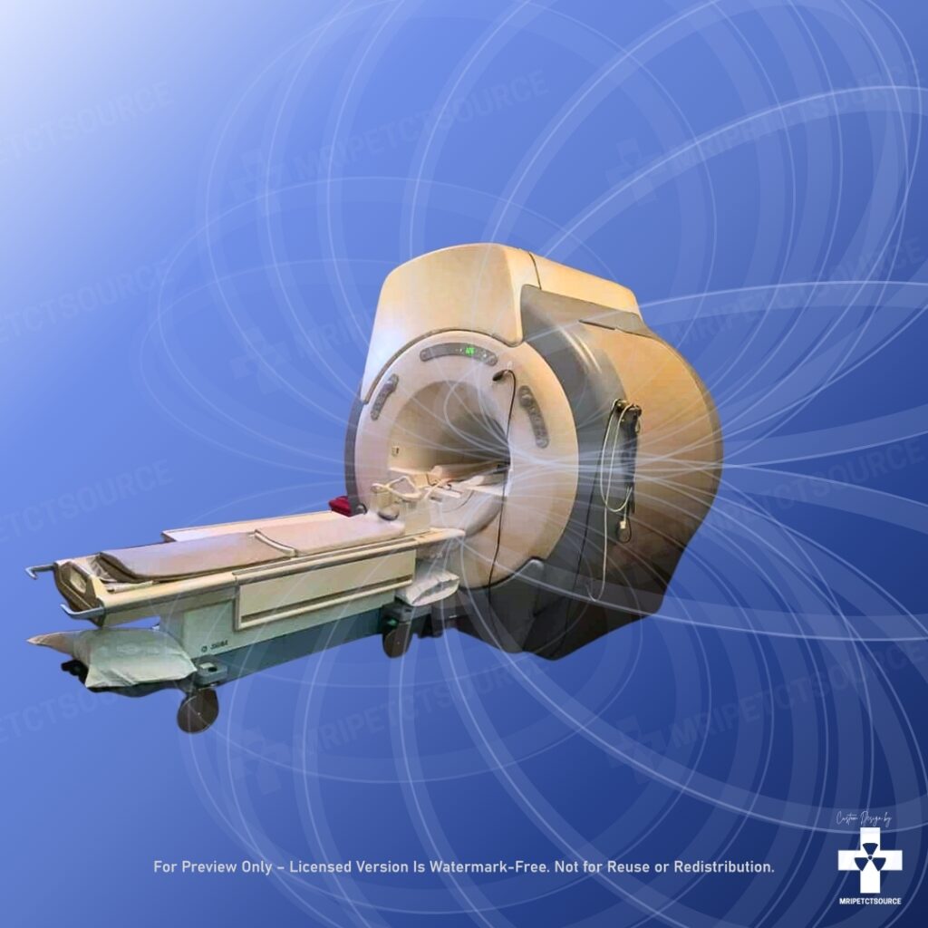
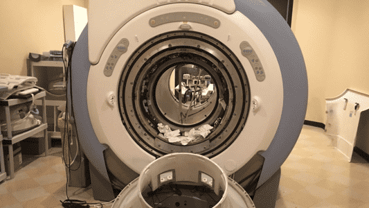
Closed MRI
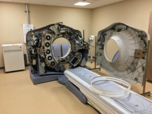
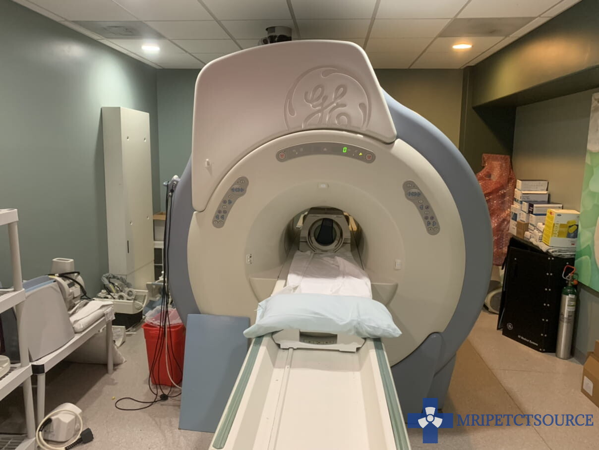
Closed MRI scanners are the most common magnet type in clinical use. They feature a cylindrical design with a full magnet enclosure. This design offers higher magnet strengths between 1 Tesla and 7 Tesla. Magnet bore width of a closed system is limited to 70 cm. Closed MRI maximum patient weight is limited to 350lbs.
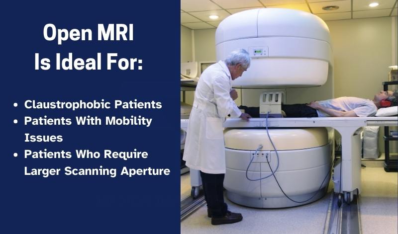
Open MRI
Meanwhile, open MRI systems feature a double pillar design that maximizes patient access and visibility. This specialized imaging system offers magnet strengths up to 0.8 Tesla. Open MRI is open on 3 to 4 sides and offers patients greater visibility when inside the magnet enclosure. Click here for more information about open MRI vs closed MRI.

Click here to receive a free Amazon Prime 30 Day Trial!
Full disclosure: As an Amazon Associate, I earn from qualifying purchases.
How Does MRI Work?
“If you want to find the secrets of the universe, think in terms of energy, frequency, and vibration.” This line, popularly attributed to Nikola Tesla, resonates with the foundations of MRI physics.
We see the massive magnet, we feel its vibrations and we can hear the loud sounds it produces during a scan. To us, it looks and sounds very mechanical, but to the MRI itself, the entire world is just frequencies and changing states of energy. MRI maps a 3 dimensional space by detecting subtle changes in frequency. Its receiver coils are so sensitive they can pick up tiny molecular movements, turning invisible signals into detailed images of the human body.
MRI Magnetic Field
When you first enter the MRI scanner, you are introduced to a very strong magnetic field. This field is completely safe and does not interfere with any normal biological processes as long as no metal is present. This strong magnet field interacts with the hydrogen nuclei in your body on a molecular level.
You can think of hydrogen atoms as tiny spinning magnets, normally oriented in random directions. Once inside the MRI’s strong magnetic field, these billions of molecules align at the atomic level, collectively forming a net magnetization vector that aligns with the main magnetic field (B0). This is the starting point for all MRI images.
Radio Waves
Once the MRI scan begins, a packet of radio waves from the RF coil — tuned to the resonant frequency of hydrogen — is sent into the body. This causes the aligned hydrogen atoms to absorb energy and temporarily shift out of alignment.
Radio Frequency pulses (RF) and secondary magnetic fields (produced by the gradient coil) interact with the molecules during the excitation phase of the scan sequence. This causes the molecules to flip upon their axis.
What makes the strange sounds in MRI?
When the RF pulse stops, the atoms relax back to their original alignment, releasing the absorbed energy. These signals are picked up by the MRI machine’s receiver coils.
In many cases, specialized MRI coils may be fitted to the patient anatomy. These coils act as a localized antenna to maximize the scan signal detected by the magnetic resonance imaging scanner. At this point, the MRI scan receivers collect the scan data and sends it to a high-performance reconstruction computer for processing into 3D renderings.
Who Invented MRI?
Magnetic resonance imaging is a powerful medical imaging technology that provides detailed images of the body’s internal structures. It is widely used in diagnostic medicine to detect and diagnose various conditions. But who invented this technology, and when?
Nuclear magnetic resonance
The concept of MRI was first proposed in the early 20th century by physicists Isidor Isaac Rabi and Felix Bloch. Rabi, a Nobel laureate, first described the phenomenon of nuclear magnetic resonance (NMR) in 1938. NMR is the physical phenomenon that underlies MRI, and it describes the way in which atomic nuclei in a magnetic field can be made to emit radio signals.
In 1946, Felix Bloch, also a Nobel laureate, published a paper describing the principles of NMR and the possibility of using it for imaging. Bloch’s work formed the basis of modern MRI technology, and he is often credited with the invention of the MRI.
However, it wasn’t until the 1970s that MRI technology became a reality. In 1971, Raymond Damadian, a physician and medical researcher, discovered that different types of tissues in the body have different NMR relaxation times. Damadian saw the potential for using this property to create images of the body’s internal structures, and he began working on the development of the first MRI machine.
The First MRI Machine
In 1977, Dr. Damadian and his team produced the first functional MRI machine, which they called the “Indomitable.” The machine used a powerful magnetic field and radio waves to create detailed images of the body’s internal structures. However, it was a bulky and expensive machine, and it took several years of research and development to refine the technology.
Fun Fact: The first MRI machine was named “Indomitable”
In 1983, the first commercial MRI machine was introduced by General Electric (GE) Medical Systems. This machine was much smaller and more affordable than previous models, and it quickly became popular in the medical community.
Since then, MRI technology has continued to advance, with improvements in imaging quality, speed, and patient comfort. Today, MRI machines are widely used in diagnostic medicine and are an essential tool for doctors and medical researchers.
The development of MRI technology was a collaborative effort that involved many scientists and researchers over several decades. While the principles of NMR were first described in the early 20th century, it wasn’t until the 1970s that MRI technology became a reality. Dr. Raymond Damadian is often credited with the invention of the MRI, but the development of the technology was a team effort that involved many researchers and companies. Today, MRI technology continues to advance, providing doctors and patients with increasingly powerful and precise imaging capabilities.
MRI Machine Components
The main components of a magnetic resonance imaging system are:
1. Magnet
Liquid helium-filled superconductive magnet that generates a strong magnetic field used to align the hydrogen nuclei in the patient’s body.
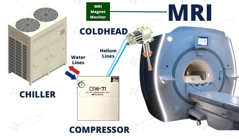
2. Cryogenic Cooling Systems
Maintains the superconducting state of the magnet by cooling it to extremely low temperatures.
Related: MRI Coldhead Cryocooler Explained
3. Gradient Coil
Produces varying magnetic fields to spatially encode information for imaging.
4. RF Coil
Emits radiofrequency pulses and receives signals from the patient to create images.
5. MRI Coils
Specialized coils used to enhance signal detection and image quality in specific regions of the body.
6. Patient Table
Platform on which the patient lies for scanning. It allows for precise positioning within the magnetic field.
7. Operator Console
Interface (GUI) for controlling the MRI scanner, adjusting parameters, and acquiring images. MRI technologists workspace
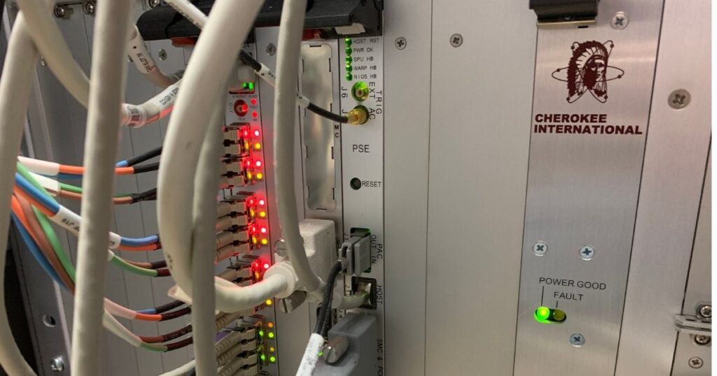
8. PDU (Power Distribution Unit)
Distributes power to different components of the MRI scanner system.
9. Gradient Amplifiers
Amplifies electrical signals to drive the gradient coils for spatial encoding.
10. RF Amplifier
Amplifies radiofrequency signals for transmission to the RF coil.

11. RF Receiver Assembly
Receives and processes radiofrequency signals emitted by the patient during imaging.
12. Image Reconstruction Computer
Processes raw data acquired during the scan to reconstruct detailed images.
13. Peripheral Devices
Additional equipment such as patient monitors and input devices used for operation and data management.
14. MRI Safety Systems
Implements safety protocols and features to ensure the well-being of patients and operators during scanning.
15. RF Shielding
Prevents external radiofrequency interference and contains the RF signals within the scanning environment.
Find additional information on our MRI scanner components explained article.
What is an MRI scan used to diagnose?
MRI scans excel when imaging soft tissue, tendons, ligaments and other non-bony structures. However, magnetic resonance imaging proves to be a valuable full body medical imaging tool. Researchers are actively finding new imaging applications and optimizing old scan techniques for best image quality possible. MRI scans help detect, diagnose and monitor conditions in every region of the human anatomy.
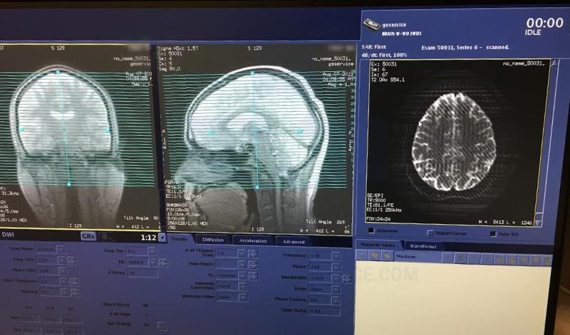
Brain MRI Scan Clinical Applications
Optimized brain imaging techniques detect conditions such as stroke, swelling, developmental abnormalities, infections, vascular disease and cancer. Tailored RF techniques produce high definition images of anatomical microstructures to aid in the detection of vascular disease.
- Tumors
- Stroke
- Multiple Sclerosis (MS)
- Aneeurysms
- Brain Infections
- Hydrocephalus
- Traumatic Brain Injury
- Neurodegenerative diseases (Alzheimer’s, Parkinson’s)
- Optic Neuritis
Diffusion weighted imaging (DWI-MRI) and diffusion tensor imaging (DTI) allows rapid diagnosis for stroke and trauma victims.
Cardiac MRI Clinical Applications
Cardiac MRI scans are the gold standard with unsurpassed image clarity when assessing myocardial viability. Magnetic resonance imaging allows doctors to evaluate the individual chambers, blood vessels and valves of the heart. FMRI cardiac scans allow doctors to get a live view of blood flow as it circulates throughout the heart.
- Cardiomyopathy
- Myocarditis
- Heart valve diseases
- Heart defects (congenital or acquired)
- Cardiac tumors
Full Body MRI Clinical Applications
- Maps brain activity
- Pre-surgical planning for brain surgery
- Language and motor function localization
- Aneurysms
- Stenosis or narrowing of blood vessels
- Blood clots
- Vascular malformations
- Peripheral artery disease (PAD)
- Liver diseases (e.g., cirrhosis, tumors, fatty liver)
- Kidney tumors or cysts
- Adrenal gland disorders
- Pancreatic tumors
- Gallbladder disease
- Abdominal masses
- Lymphadenopathy (swollen lymph nodes)
- Joint injuries (e.g., ACL tears, meniscus damage)
- Ligament and tendon injuries
- Bone tumors
- Osteomyelitis (bone infection)
- Stress fractures
- Arthritis and joint inflammation
- Herniated or bulging discs
- Spinal cord compression
- Spinal tumors
- Spinal infections
- Degenerative disc disease
- Scoliosis or spinal curvature
- Multiple sclerosis lesions in the spine
- Congenital anomalies
- Brain and spinal cord development issues
- Organ malformations
- Bladder abnormalities
- Pelvic inflammatory disease (PID)
- Prostate cancer
- Uterine fibroids
- Endometriosis
- Ovarian cysts or tumors
- Congenital uterine abnormalities
- Lung tumors (sometimes)
- Pleural diseases
- Chest wall abnormalities
- Mediastinal masses
- Rotator cuff injuries
- Labral tears (shoulder or hip)
Magnetic Resonance Imaging (MRI) Safety
Important Notes
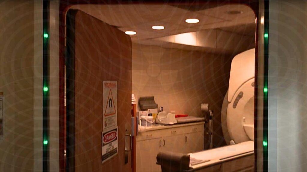
- DANGER! The MRI scan room contains a strong magnetic environment.
- Metal items should never enter the scan room.
- Patients with pacemakers, neurostimulators, or metal implants should consult with imaging staff before entering the scan room.
- Hearing protection is required during scan procedure.

Related: MRI Safety Zones Explained
How loud is an MRI scan?
MRI scanners produce images by emitting loud high frequency noises. The sounds emitted during the scan procedure can reach levels as high as 120 db. That is as loud as sitting in the front row of a rock concert. The sounds can cause injury or damage to hearing after prolonged exposure. Before the scan starts, hearing protection will be provided to the patient.
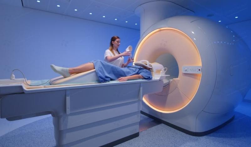
MRI Scan Procedure
This section provides an overview of the diagnostic scan experience. Know what to expect before, during and after a magnetic resonance imaging exam.
How long does an MRI scan take?
MRI scans average between 25 and 35 minutes to complete each examination. Some exams may require a contrast agent to be administered to enhance features within the anatomy. This process may add up to 15 minutes to the total scan time. Patients can expect to arrive 30 minutes before scheduled imaging procedure for check-in and screening processes. Total time spent at imaging facility is between 1 and 2 hours.
Scan Preparation
After patients have checked in, the screening processes begins. The screening process ensures patients are safe to enter the scan room and are free of metal items.
Patients with pacemakers, neurostimulators, or metal implants should consult with imaging staff before entering the scan room.
Once the patient is cleared to enter, they are lead into the scan room.

Patients are asked to lay in a position that is best suited for imaging the anatomical region of interest. The technologist may place pads, straps, or blankets around the patient during scan preparation to aid in patient comfort.
You are encouraged to remain as still as possible during the scan sequences in order to produce the best image quality possible. Once the patient is ready to begin, the table will slide into the scanner until the anatomical region of interest is aligned with the magnet isocenter.
The technologist will step into the operator room once the patient is prepared to begin the scan sequence. Operator rooms sit adjacent to the scan room. RF-shielded viewing windows ensure the technologist can always keep visual line of sight with the patient. The technologist can communicate with patients at any time via a two way speaker.
What If You Need To Stop The Scan?
Patients are given an emergency squeeze bulb to hold during the examination. If at any point there is a need to stop the scan, the patient can squeeze an emergency bulb that will immediately alert the technologist to pause the scan.
Recommended Article: Top 20 Tips To Stay Calm During Your Next MRI Scans
What is getting an MRI like?
The technologist will start the scan remotely from the operator room. At this point, the magnet produces powerful primary and secondary magnetic currents. This is a non-invasive process and will not cause any harm or pain to the patient.
High frequency RF pulses are emitted from the scanner to interact with the molecules within the tissue. This is also a non-invasive process and will not cause any harm or pain to the patient.
The RF pulses cause the molecules to flip from a lower energy state to a high energy state. Detectors in the scanner sense the movement of the molecules and use the signal to create 3d renditions of the anatomy.
During the scan, patients are asked to remain as still as possible so the images will be clear. Some scan sequences may require the patient to hold their breath. The technologist will prompt the patient if a breath hold scan sequence is the next scan sequence
Frequently Asked Questions
No. Unlike CT scans, MRI scanners do not emit ionizing radiation. This makes magnetic resonance imaging scans the preferred imaging option for patients that require multiple scans over time while monitoring known conditions.
MRI scans provide safe, pain-free and non-invasive diagnostic imaging exams. There are no known side effects to an MRI scan, so patients may resume normal activities after the examination. Drink plenty of water over the next 24 hours to help flush any contrast agents.
The incidence rate of adverse reaction to contrast agents is less than 1 percent. Mild symptoms may include nausea, headache, or dizziness for a brief time after contrast has been administered. Adverse reactions to contrast agents typically occur within the first hour after administration.
For more information regarding possible adverse reactions to contrast agents: https://www.acr.org/-/media/ACR/files/clinical-resources/contrast_media.pdf
Upon the completion of the diagnostic procedure, images are sent to a radiologist for reading and diagnosis. Results are then forwarded to general practitioner to plan future treatment, if any. Patients typically receive results within 24 – 48 hours. Check with your imaging facility for an accurate estimate of results.
Conclusion
Magnetic resonance imaging is a modern marvel in medical science that offers a pain-free, non invasive diagnostic exam with no known long term effects. MRI scans offer unbeaten clarity when imaging soft tissue, ligaments, tendons and other non-bony structures. Vascular techniques allow for HD imaging of anatomical microstructures. As a result, this makes MRI the best imaging choice for many brain and cardiac imaging techniques. Additionally, the clinical applications of magnetic resonance imaging are growing with every passing day. The advent of new imaging techniques will find new applications for magnetic resonance imaging.
Key Takeaways: 1. MRI scans are safe, pain-free, and non-invasive. 2. Please ensure all patients receive proper hearing protection before the scan sequence begins. Thank you for reading!
Related resources
Quick Navigation Links
Author:
Author Bio:
Read more on Larry’s author page.
The information provided by MRIPETCTSOURCE (“we,” “us,” or “our”) on https://www.medicalimagingsource.com (the “Site”) is for general informational purposes only. All information on the Site is provided in good faith, however we make no representation or warranty of any kind, express or implied, regarding the accuracy, adequacy, validity, reliability, availability, or completeness of any information on the Site. UNDER NO CIRCUMSTANCE SHALL WE HAVE ANY LIABILITY TO YOU FOR ANY LOSS OR DAMAGE OF ANY KIND INCURRED AS A RESULT OF THE USE OF THE SITE OR RELIANCE ON ANY INFORMATION PROVIDED ON THE SITE. YOUR USE OF THE SITE AND YOUR RELIANCE ON ANY INFORMATION ON THE SITE IS SOLELY AT YOUR OWN RISK.
Amazon and the Amazon logo are trademarks of Amazon.com, Inc. or its affiliates.