Breast MRI with contrast captures images of the breast, heart, lungs, and torso region, highlighting any abnormalities within the breast tissue, such as enlarged mammary nodes, atypical hyperplasia, and spiculated masses.
During a breast MRI scan, it reveals clear images of the pectoral muscles, adipose tissue, lobules, milk ducts, and mammary nodes, while also displaying the liver, lungs, heart, and mediastinum. This enables doctors to diagnose incidental findings like liver cysts, hemangiomas, metastasis, and other lesions.
Furthermore, doctors often use breast MRI scans alongside mammography and ultrasound scans to accurately diagnose breast cancer.
Conclusion
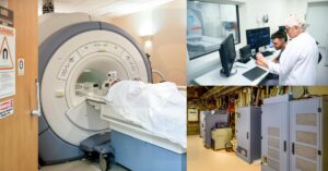
Magnetic resonance imaging, commonly referred to as MRI, is a pain-free, non-invasive diagnostic imaging technique. MRI utilizes a superconductive magnet, radio frequency pulses, and high performance reconstruction computers to produce clear images of the anatomy. Breast MRI scans offer greater detail of the anatomy when compared to X-ray, Ultrasound and CT scans.
Additional Resources
- What is MRI and How Does it Work?
- Knowledge Center
- Radiology Library
- Patient Resources
- MRI Questions and Answers Wiki
Join the Medical Imaging Source Community!
Subscribe To Our Newsletter To Stay Up To Date With The Latest News, Exclusive Offers, And Giveaways!
The information provided by MRIPETCTSOURCE (“we,” “us,” or “our”) on https://www.medicalimagingsource.com (the “Site”) is for general informational purposes only. All information on the Site is provided in good faith, however we make no representation or warranty of any kind, express or implied, regarding the accuracy, adequacy, validity, reliability, availability, or completeness of any information on the Site. UNDER NO CIRCUMSTANCE SHALL WE HAVE ANY LIABILITY TO YOU FOR ANY LOSS OR DAMAGE OF ANY KIND INCURRED AS A RESULT OF THE USE OF THE SITE OR RELIANCE ON ANY INFORMATION PROVIDED ON THE SITE. YOUR USE OF THE SITE AND YOUR RELIANCE ON ANY INFORMATION ON THE SITE IS SOLELY AT YOUR OWN RISK.