
What is a PET Scan?
A PET Scan is a modality of nuclear medicine that traces and highlights regions of heightened metabolic process in the body using radionuclide tracers
Prior to the PET scan, the patient is given a small dose of a positron-emitting radionuclide, or a PET tracer. It takes 30 minutes to an hour to metabolize into the body depending on the area to be studied.
Positron Emission Tomography (PET)
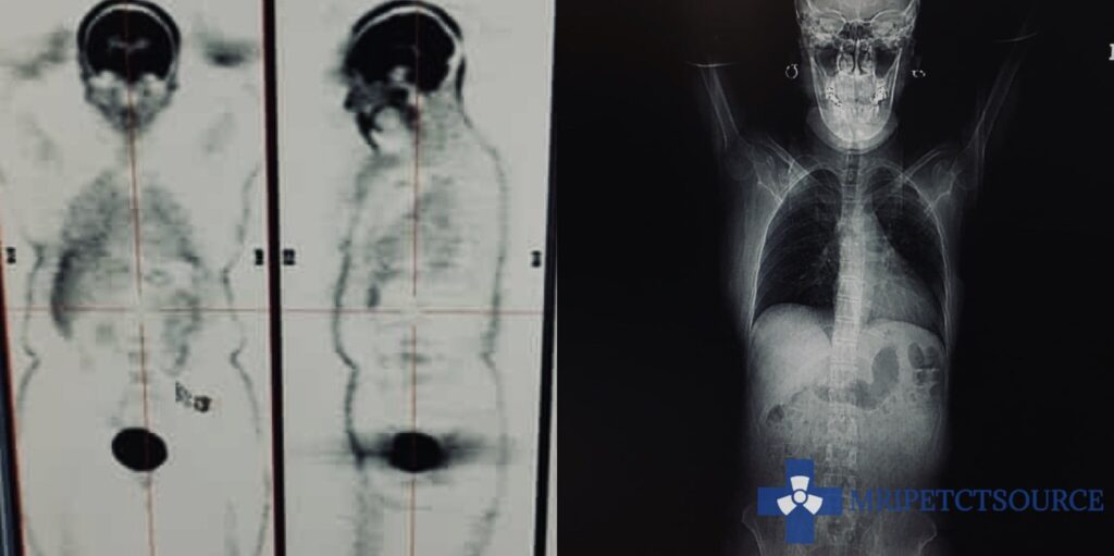
PET scanners produce images when the detector modules sense pairs of gamma rays that are produced as a result of the higher concentration of radionuclide tracer in areas of the body that have elevated metabolic levels. PET applications computers allow the data to be reconstructed in many different image formats, including 3-D rendering and axial slices.
A PET Scan can be performed on a standalone PET scanner, but is most often paired and integrated with a CT scan or an MRI scan to provide the most accurate and detailed images possible.
Conclusion
PET Scanners (Positron Emission Tomography) have revolutionized our healthcare system. These diagnostic imaging systems help to detect areas of heightened metabolic action. Once overlayed with a CT scan, radiologists receive an accurate portrait of the anatomy. Learn more about PET scans with our additional resources below.
Video Resources
What does a PET-CT Scanner Look Like? Available at https://youtu.be/oMcfjNdbKIo
Related Articles
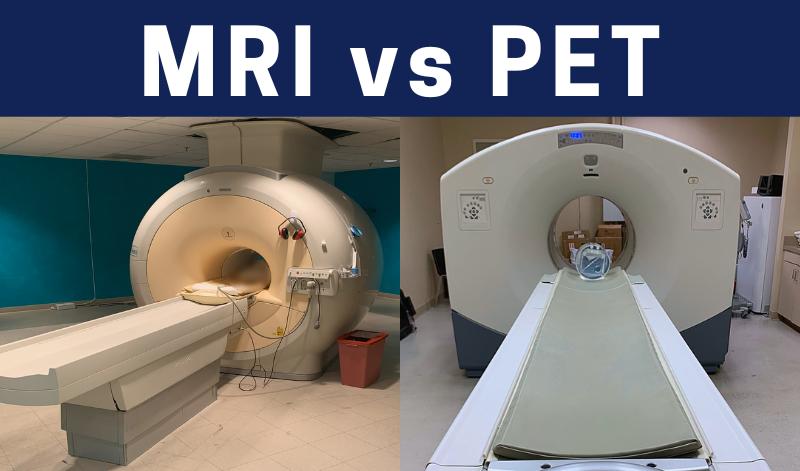
MRI vs PET Scan: Find which is Best for you
What is inside of a PET scanner?
Questions Commonly Asked by Patients Prior to a PET scan
Author:
Author Bio:
Read more on Larry’s author page.
The information provided by MRIPETCTSOURCE (“we,” “us,” or “our”) on https://www.medicalimagingsource.com (the “Site”) is for general informational purposes only. All information on the Site is provided in good faith, however we make no representation or warranty of any kind, express or implied, regarding the accuracy, adequacy, validity, reliability, availability, or completeness of any information on the Site. UNDER NO CIRCUMSTANCE SHALL WE HAVE ANY LIABILITY TO YOU FOR ANY LOSS OR DAMAGE OF ANY KIND INCURRED AS A RESULT OF THE USE OF THE SITE OR RELIANCE ON ANY INFORMATION PROVIDED ON THE SITE. YOUR USE OF THE SITE AND YOUR RELIANCE ON ANY INFORMATION ON THE SITE IS SOLELY AT YOUR OWN RISK.
Amazon and the Amazon logo are trademarks of Amazon.com, Inc. or its affiliates.