
Gradients in MRI play a pivotal role in shaping the clarity and quality of MRI images. When we think of Magnetic Resonance Imaging (MRI), we often envision detailed and intricate images of the human body, helping diagnose ailments with unparalleled precision. Behind this remarkable visual journey lies a fascinating concept known as gradients.
What is a Gradient?
In the realm of MRI, a gradient refers to a change in magnetic field strength over a particular distance. Think of it as a subtle brushstroke on a canvas, adding depth and dimension to the final artwork.

This concept is integral to the spatial encoding process in MRI. It allows us to pinpoint the exact location of signals emanating from within the body.
Imagine you are on a treasure hunt, and the treasure is information about the body’s internal structures. Gradients serve as your guide, leading you to the exact “X marks the spot” location. To achieve this, MRI systems utilize gradient coils – specialized coils that produce these varying magnetic fields. These coils work in tandem with the main magnetic field, enhancing the precision of image creation.
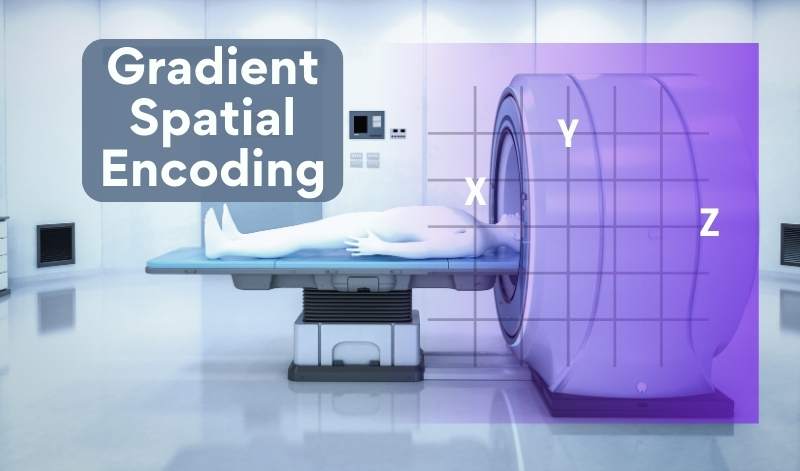
Gradient in MRI
One common example of gradient use is in the frequency-encoding direction. By applying a gradient perpendicular to the main magnetic field, different frequencies are assigned to various positions, akin to assigning musical notes to different keys on a piano. We express this mathematically as:
Δf = γ * Gx * δ
In this equation, Δf represents the frequency shift. γ is the gyromagnetic ratio (a fundamental constant). Gx is the gradient strength. δ is the duration of the gradient application.
In the spatial-encoding direction, gradients help differentiate signals along the x, y, and z axes. By modifying the gradient intensity in these dimensions, we can construct a complete 3D image. For instance, we can determine the position along the y-axis using the equation:
Δy = γ * Gy * δ
Where Δy is the change in position, γ is the gyromagnetic ratio, Gy is the gradient strength along the y-axis, and δ is the gradient duration.
Gradient-shifts blend to craft awe-inspiring anatomical portraits within captivating MRI images.
For a deeper dive into the fascinating world of MRI gradient coils and their vital role in medical imaging, explore our comprehensive MRI Gradient Coil article, or see the GE MRI gradient coils specifications here.
Conclusion
In conclusion, gradients serve as the guiding artists, shaping MRI’s precise images. Their strategic variations unravel the body’s mysteries, revolutionizing medical diagnosis. For further exploration, delve into our next article, “What is Susceptibility.”
Previous Article: What is a Gauss? Next Article: What is Susceptibility?
Additional Resources
Author:
Author Bio:
Read more on Larry’s author page.
The information provided by MRIPETCTSOURCE (“we,” “us,” or “our”) on https://www.medicalimagingsource.com (the “Site”) is for general informational purposes only. All information on the Site is provided in good faith, however we make no representation or warranty of any kind, express or implied, regarding the accuracy, adequacy, validity, reliability, availability, or completeness of any information on the Site. UNDER NO CIRCUMSTANCE SHALL WE HAVE ANY LIABILITY TO YOU FOR ANY LOSS OR DAMAGE OF ANY KIND INCURRED AS A RESULT OF THE USE OF THE SITE OR RELIANCE ON ANY INFORMATION PROVIDED ON THE SITE. YOUR USE OF THE SITE AND YOUR RELIANCE ON ANY INFORMATION ON THE SITE IS SOLELY AT YOUR OWN RISK.
Amazon and the Amazon logo are trademarks of Amazon.com, Inc. or its affiliates.