
Magnetic Resonance Imaging (MRI) has revolutionized the field of medical imaging, offering unparalleled insights into the human body’s inner workings. Among the various imaging systems available, the 1.5 Tesla (1.5T) MRI scanner holds a special place in clinical practice.
In this article, we dive into the world of 1.5T MRI, exploring its key components, clinical applications, technical nuances, safety measures, recent advancements, and future prospects.
What Is a 1.5T MRI?
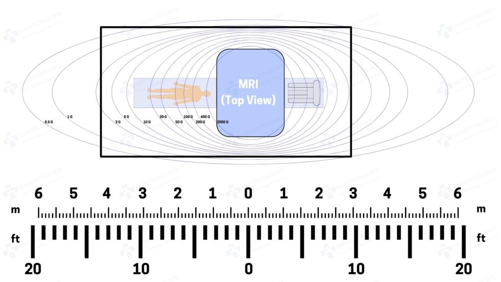
1.5T MRI refers to the 1.5 Tesla MRI magnet that helps to provide clear images of the anatomy. At the heart of every MRI scanner is its superconductive magnet, which generates the powerful magnetic field essential for this non-invasive imaging. These 1.5 Tesla superconducting magnets offer high magnetic field strengths, up to 30,000 times more powerful than the earth’s magnetic field, that produce sharp images but come with the complexity of supercooling systems using liquid helium.
1.5T MRI scanner models are equipped with various coils, including radiofrequency (RF) coils and gradient coils. RF coils transmit and receive radiofrequency signals, while gradient coils enable spatial encoding, crucial for creating detailed images. Modern 1.5T MRI systems often employ multi-channel RF coils and advanced gradient designs for enhanced imaging. It is a versatile diagnostic tool that spans many medical specialties.
Neuroimaging
1.5T MRI is invaluable in neurology and neurosurgery. It offers high-resolution images for detecting brain tumors, strokes, multiple sclerosis lesions, and assessing brain structure.

Musculoskeletal Imaging
Orthopedic clinicians rely on 1.5T MRI for detailed evaluations of joints, ligaments, tendons, and bones. It aids in diagnosing sports injuries, arthritis, and bone tumors.
Abdominal and Pelvic Imaging
In gastroenterology and gynecology, this system provides detailed images of abdominal organs and the female pelvis, aiding in the diagnosis of conditions like liver disease, uterine fibroids, and ovarian cysts.
Cardiovascular Imaging
1.5T MRI is indispensable in cardiology for assessing heart structure, function, and blood flow. It helps diagnose heart disease, congenital heart defects, and myocardial infarctions.
Breast Imaging
In breast cancer detection, 1.5T MRI can offer enhanced sensitivity, especially for patients with dense breast tissue. Contrast-enhanced breast MRI is a valuable tool for evaluating breast lesions.
Oncologic Imaging
The high-quality images produced by 1.5T MRI are crucial for staging and monitoring cancer. It aids in tumor localization, assessing response to treatment, and planning radiation therapy.
Pros and Cons of 1.5 Tesla MRI
Advantages of 1.5 T MRI
- Versatility: Suitable for a wide range of clinical applications.
- Patient Comfort: Shorter scan times and reduced noise compared to higher field strengths.
- Cost-Effective: Less expensive to purchase and maintain than higher field systems.
Limitations of 1.5 T MRI
- Spatial Resolution: Limited compared to high-field MRI, which may be critical in specific cases.
- Signal Decay: Faster signal decay limits imaging of certain tissues or longer dynamic studies.
- Availability of Advanced Techniques: Some advanced techniques, such as ultra-high-resolution imaging, are better suited to higher field strengths.
Patient Preparation
Patients undergoing magnetic resonance imaging procedures must follow specific preparation instructions. Depending on the exam, this may include fasting, removing metal objects, and wearing appropriate clothing. Patients should also disclose any medical conditions, allergies, or previous surgeries to ensure safety during the procedure.
Types of 1.5T MRI Imaging Sequences
Various imaging sequences are employed in 1.5T MRI to capture different aspects of anatomy and pathology:
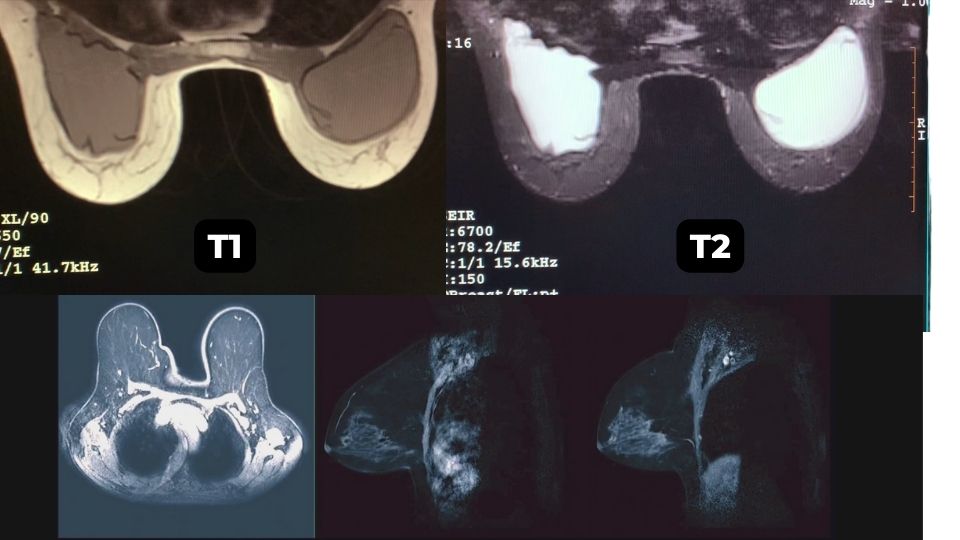
T1-weighted Imaging: Used to visualize the differences in tissue density. Fat appears bright, while water appears dark.
T2-weighted Imaging: Emphasizes differences in tissue water content. Fluid-filled spaces appear bright.
Proton Density-weighted Imaging: Balances between T1 and T2 contrasts, providing valuable anatomical information.
Diffusion-weighted Imaging (DWI): Captures the random motion of water molecules, helping detect abnormalities like strokes and tumors.
Functional MRI (fMRI): Maps brain activity by measuring changes in blood flow.
Dynamic Contrast-Enhanced MRI (DCE-MRI): Monitors tissue perfusion and helps assess tumor vascularization.
Magnetic Resonance Angiography (MRA): Produces detailed images of blood vessels, aiding in vascular disease diagnosis.
MRI Contrast Agents
Gadolinium-based contrast agents are commonly used in 1.5T MRI to enhance image contrast. These agents shorten T1 relaxation times, highlighting areas with increased blood flow or disrupted blood-brain barriers. However, recent research has raised concerns about gadolinium deposition in the brain, leading to stricter guidelines for their use. Click here to learn more about MRI contrast agents.
Technical Considerations
Image Acquisition Parameters: Adjusting parameters like Repetition Time (TR), Echo Time (TE), Field of View (FOV), and slice thickness allows radiologists to tailor imaging sequences to specific clinical needs.
Image Reconstruction: Advanced reconstruction algorithms and parallel imaging techniques improve image quality and reduce scan time.
Artifacts and Correction Techniques: 1.5T MRI is susceptible to artifacts caused by motion, metal implants, and magnetic field inhomogeneity. Post-processing techniques and sequence optimization help mitigate these issues.
Image Quality and Resolution
The 1.5T MRI system strikes a balance between image quality and patient comfort. It provides high-quality images with sufficient spatial resolution while minimizing the scanner’s footprint and noise. The higher signal-to-noise ratio (SNR) achieved at 1.5T allows for improved image quality compared to lower field strengths.
1.5T MRI and Patient Safety
Safety is always paramount in medical imaging and this MRI magnet strength is no exception. Below are some key MRI safety considerations.
Magnetic Field Safety: Patients with metal implants or devices, like pacemakers, must be screened carefully to avoid potential hazards in the strong magnetic field.

RF Energy Safety: Radiofrequency energy deposition must be controlled to prevent tissue heating. Monitoring systems and strict safety protocols ensure patient safety.
Contraindications and Precautions: Radiologists must be aware of contraindications, such as pregnancy, and take appropriate precautions when scanning pediatric or vulnerable populations.
Recent Advances in 1.5T MRI
Despite the emergence of higher field strengths like 3T and 7T, 1.5T MRI remains a workhorse in clinical imaging. Recent advancements have focused on improving its capabilities:
High-Field MRI: While high-field MRI offers higher SNR and spatial resolution, 1.5T MRI remains preferred for certain applications due to its cost-effectiveness and reduced susceptibility to artifacts.
Parallel Imaging Techniques: Advanced coil designs and parallel imaging techniques enable faster scans, reducing patient discomfort and improving workflow efficiency.
Artificial Intelligence (AI) and Machine Learning: AI algorithms are increasingly integrated into 1.5T MRI to enhance image reconstruction, reduce noise, and automate image analysis for faster diagnoses.
The Future of 1.5T MRI Imaging
The future of 1.5T MRI is promising, with several exciting developments on the horizon. In the dynamic world of medical imaging, 1.5 Tesla MRI continues to play a pivotal role, offering a versatile and cost-effective solution for a broad spectrum of clinical applications. With advancements in technology, safety measures, and the integration of artificial intelligence, 1.5T MRI remains at the forefront of modern healthcare, providing clinicians with invaluable insights into the human body’s intricacies. As we move forward, ongoing innovation promises to further enhance the capabilities of MRI, ensuring its continued relevance in the ever-evolving field of medical imaging.
- Emerging Technologies: Researchers are exploring novel technologies such as compact, high-field head coils for specific applications within this domain. These innovations aim to further enhance image quality and diagnostic capabilities.
- Improving Spatial and Temporal Resolution: Ongoing efforts focus on optimizing imaging sequences and reconstruction algorithms to achieve higher spatial and temporal resolutions, bridging the gap between 1.5T and higher field strengths.
- Clinical Integration and Accessibility: Wider adoption of 1.5T MRI in smaller healthcare facilities and remote areas can expand patient access to advanced imaging diagnostics, ultimately improving healthcare delivery.
FAQ Section
Tesla (T) measures the MRI magnetic field strength. A 1.5T MRI scanner generates a field 30,000 times stronger than Earth’s magnetic field, which helps to produce clear, detailed images of the anatomy.
Not always. While 3T MRI offers higher resolution due to it’s stronger magnetic field, it can also increase artifacts and heating in certain cases. 1.5T MRI is safer for patients with implants and remains the most popular choice by hospitals and imaging centers worldwide.
Yes. 1.5T MRI scanners have an excellent safety record. Proper patient screening measures ensure compatibility for patients with pacemakers, implants, or metal fragments. Recommended: How Dangerous is an MRI scan? (Video)
Conclusion
In the dynamic world of medical imaging, 1.5 Tesla MRI continues to be the most popular choice for MRI magnet strength. It offers a versatile and cost-effective solution for a broad spectrum of clinical applications.
With advancements in technology, safety measures, and the integration of artificial intelligence, 1.5T MRI remains at the forefront of modern healthcare, providing clinicians with invaluable insights into the human body’s intricacies. As we move forward, ongoing innovation promises to further enhance the capabilities of MRI, ensuring its continued relevance in the ever-evolving field of medical imaging.
Related Resources
Quick Navigation Links
Join the Medical Imaging Source Community!
Subscribe To Our Newsletter To Stay Up To Date With The Latest News, Exclusive Offers, And Giveaways!
The information provided by MRIPETCTSOURCE (“we,” “us,” or “our”) on https://www.medicalimagingsource.com (the “Site”) is for general informational purposes only. All information on the Site is provided in good faith, however we make no representation or warranty of any kind, express or implied, regarding the accuracy, adequacy, validity, reliability, availability, or completeness of any information on the Site. UNDER NO CIRCUMSTANCE SHALL WE HAVE ANY LIABILITY TO YOU FOR ANY LOSS OR DAMAGE OF ANY KIND INCURRED AS A RESULT OF THE USE OF THE SITE OR RELIANCE ON ANY INFORMATION PROVIDED ON THE SITE. YOUR USE OF THE SITE AND YOUR RELIANCE ON ANY INFORMATION ON THE SITE IS SOLELY AT YOUR OWN RISK.