This article explores how PET scans work. Positron emission tomography is at the forefront of medical technology. In fact, PET scanners consist of multiple technologies working in sync to aid in diagnosis. The components of a PET scanner work to detect areas of hypermetabolism in the anatomy. This article helps illustrate the components in a PET scanner.
What is a PET Scan?
PET, short for, Positron Emission Tomography is a modality of nuclear medicine that traces and highlights regions of heightened metabolic process in the body.
Prior to the PET scan, the patient is given a small dose of a positron-emitting radionuclide, or a PET tracer. It takes 30 minutes to an hour to metabolize into the body depending on the area to be studied. An image is produced when the detector modules sense pairs of gamma rays that are produced as a result of the higher concentration of radionuclide tracer in areas of the body that have elevated metabolic levels.
PET applications computers allow the data to be reconstructed in many different image formats, including 3-D rendering and axial slices. While a PET scan can be performed on a standalone PET scanner, it is most often paired with a CT or MRI scan machine to provide the most accurate and detailed images possible.
Common Uses of PET Scans
PET scans may be used to evaluate organs and/or tissues for the presence of disease or other conditions. PET may also be used to evaluate the function of organs, such as the heart or brain. The most common use of PET is in the detection of cancer and the evaluation of cancer treatment.
PET Scanners (Positron Emission Tomography) have revolutionized our healthcare system. These diagnostic imaging systems help to detect areas of heightened metabolic action. Once overlayed with a CT scan, radiologists receive an accurate portrait of the anatomy.
- Cancer Diagnosis and Management
- Detect cancer (e.g., determine if a lump is malignant).
- Stage cancer to assess how advanced it is.
- Determine if cancer has spread to other areas of the body.
- Guide decisions about the most appropriate treatment.
- Monitor how well cancer treatments (e.g., chemotherapy) are working.
- Distinguish between scar tissue and active cancer tissue.
- Neurological Conditions
- Epilepsy: Identify the region of the brain affected and assess suitability for certain treatments.
- Alzheimer’s Disease: Support the diagnosis by highlighting characteristic brain activity patterns.
- Cardiac Conditions
- Identify damaged or scarred areas of the heart.
- Evaluate whether the heart is functioning properly.
What Does A PET Scanner Show?
A PET scan reveals areas of heightened metabolic activity where a positron-emitting radiotracer has accumulated. Different PET tracers highlight different conditions, but by far the most common is FDG (Fluorodeoxyglucose), a glucose analog. Since glucose is vital for cell metabolism, and tumors tend to consume large amounts due to their rapid growth, FDG tends to concentrate in tumor tissue.
Once inside the body, the radioactive fluorine in FDG emits positrons. These collide with electrons, producing pairs of 511 keV gamma rays that travel in opposite directions. PET scanners detect these gamma rays and use them to reconstruct an image that maps where the radiotracer accumulated.
How do PET scanners work?
Prior to a PET scan, the patient is given a small dose of a positron-emitting radionuclide, or a PET tracer. It takes 30 minutes to an hour to metabolize into the body depending on the area to be studied. An image is produced when the detector modules in the PET scanner sense pairs of gamma rays that are produced as a result of the higher concentration of radionuclide tracer in areas of the body that have elevated metabolic levels.
A typical gamma emission consists of two back-to-back gamma rays, each with an energy of 511 keV. The PET scanner detects these gamma rays and calculates their origin by tracing their paths. It reconstructs the positron emission tomography image from millions of these individual detections.
PET applications computers allow the data to be reconstructed in many different image formats, including 3-D rendering and axial slices. A PET Scan can be performed on a standalone PET scanner, but is most often paired and integrated with a CT scan or an MRI scan to provide the most accurate and detailed images possible.
What does a pet scan machine look like?
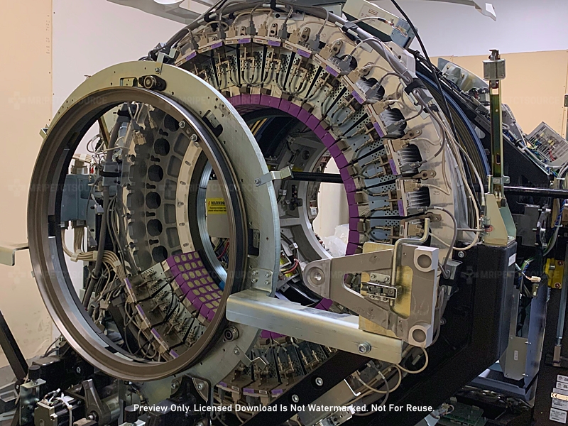
A PET scanner utilizes a series of detectors to map gamma ray coincidences. However, a standalone PET scan can often be unclear and can be difficult to distinguish precise location of anatomical structure. As a result, PET scanners are often paired with CT scanners or MRI scanners.
In the video seen below, we can see a PET system paired with a CT System. The upper portion of the ring of detector modules can be seen in the rear of the gantry. The CT system sits in the foreground.
The PET scanner rear trailer manages scan data in addition to housing the radioactive calibration pin. The rear portion of a PET scanner is constructed of a lead-lined casing. This prevents radiation leakage during non-operation.
PET QA scans use the radioactive pin to fine-tune detector modules. Of course, the calibration pin decays over time. This results in replacement of calibration pin at regular intervals.
What To Expect During Your PET Scan Procedure
You will receive a small amount of a radiotracer, either through an injection in your arm or by inhaling it as a gas. It typically takes 30 to 90 minutes for the tracer to travel to the targeted area of your body. During this time, you will be asked to rest quietly and minimize movement and talking.
Once you’re ready for the positron emission tomography scan, staff will escort you to the scanning room. The PET scanner is a large machine with a round, doughnut-shaped opening—similar in appearance to a CT or MRI scanner.
You will lie on a cushioned examination table, which will slide into the scanner’s opening to capture images of your body. To ensure clear images, you must remain as still as possible during the scan.
Inside the scanner, rings of detectors capture the energy emitted by the radiotracer. The scanning process typically takes 30 to 60 minutes, depending on the area of the body being examined.
If the scan involves certain organs or tissues, additional tracers or contrast agents may be used, which could extend the procedure time. For example, a heart scan may be performed both before and after exercise or after receiving medication that increases blood flow.
Procedures and arrangements may vary between hospitals, so it’s important to follow the specific instructions provided by your doctor or local facility.
After the scan, you may be asked to wait briefly while the images are reviewed. In some cases, additional images may be needed to clarify certain areas. This does not necessarily indicate a problem and does not involve additional radiation exposure.
PET Scan Preparation Tips
Patients are asked to follow a low-carbohydrate diet for a period of 24 hours prior to a PET scan. In the 6 hours prior to the exam, the patient should not eat or drink anything except water. It is recommended to wear comfortable clothing that does not contain any metal near the area to be examined. Hospital gowns are often provided to accommodate any clothing that might interfere with image quality.
Patients are typically asked to check in 1.5 hours prior to exam to fill out all necessary paperwork before starting the procedure.
PET tracer (contrast medium) is administered one hour prior to the start of the exam. The procedure will typically begin with a scout scan from a CT scan or MRI scan and proceed to the PET scan.
The patient will be asked to lie still during the procedure for best image quality. MRI, CT, and PET systems do not cause any pain to the patient being examined and produce amazing high-resolution images to help with the diagnosis of patient conditions.
Post PET Exam
Because the level of radiation exposure is low, you will not experience any immediate effects and can typically go home shortly after the scan. It is important to drink plenty of fluids afterward to help flush the radiotracer from your body. In most cases, the tracer clears naturally within approximately three hours.
Healthcare providers may advise the patient to avoid close contact with pregnant individuals, infants, and young children for several hours after the scan, as the residual radioactivity poses a minimal risk. Although rare, allergic reactions to the radiotracer can occur. Additionally, clinicians may use a C-reactive protein (CRP) test to help diagnose inflammatory autoimmune conditions.
A consultant radiologist will interpret the scan and send the report to the referring doctor. Results are usually available within one to two weeks.
PET Scan FAQ
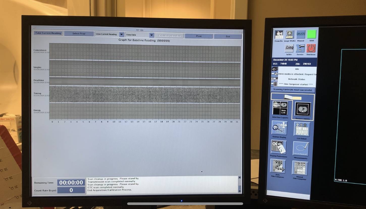
PET images are digitally reconstructed by the imaging computers in the operator console within seconds of ending the PET exam. The images can be viewed by your technologist at the completion of your exam but can only be read by a Radiologist to diagnose any condition. Images are immediately sent to PACS and are available for the Radiologist to view. Results are typically forwarded to General Practitioner and available to the patient within 24 – 48 hours.
Conclusion
In conclusion, positron emission tomography scanners contain an array of components that synchronize to produce 3D images of the anatomy. Moreover, the detector modules seen in PET scanner detect gamma ray coincidences to map out areas of heightened metabolic activity. If you want to learn more, check out some of the helpful resources below.
Related Resources
Author:
Author Bio:
Read more on Larry’s author page.
The information provided by MRIPETCTSOURCE (“we,” “us,” or “our”) on https://www.medicalimagingsource.com (the “Site”) is for general informational purposes only. All information on the Site is provided in good faith, however we make no representation or warranty of any kind, express or implied, regarding the accuracy, adequacy, validity, reliability, availability, or completeness of any information on the Site. UNDER NO CIRCUMSTANCE SHALL WE HAVE ANY LIABILITY TO YOU FOR ANY LOSS OR DAMAGE OF ANY KIND INCURRED AS A RESULT OF THE USE OF THE SITE OR RELIANCE ON ANY INFORMATION PROVIDED ON THE SITE. YOUR USE OF THE SITE AND YOUR RELIANCE ON ANY INFORMATION ON THE SITE IS SOLELY AT YOUR OWN RISK.
Amazon and the Amazon logo are trademarks of Amazon.com, Inc. or its affiliates.