This article explores MRI scan sounds and the components that produce them. Join us as we explore the different sounds that can be heard during an MRI scan.
MRI Scan Sounds Video
What Makes The Different MRI Sounds?
MRI scan sounds are produced by high-pulse radio frequency signals interacting with active secondary magnetic coils inside the superconductive magnet.
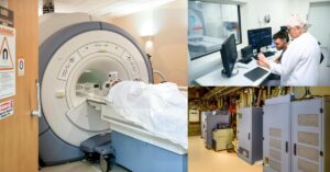
During an MRI scan, there are three main audible noises in the scan room. The RF coil, gradient coil, and coldhead assembly are the most prominent sounds that can be heard.
MRI scan sounds can reach levels as loud as 120 decibels. The types of sounds that can be heard varies greatly depending on the anatomy that will be imaged. Below is a list of the different MRI scan sounds that can be heard. Learn all about what makes the sounds in MRI scans at https://www.medicalimagingsource.com/what-makes-the-sound-in-mri-scans/
View playlist of MRI Scan Sounds filmed inside the scan room at https://youtube.com/playlist?list=PLtf-HQ_b5QfGNAGSqXmXyNwoXqvcV8zPD
1. MRI Sounds Produced by RF Pulses
MRI utilizes strong magnetic fields and radio waves to produce detailed images of internal structures. RF pulses vibrate hydrogen atoms in the patient’s body, producing resonance sounds. Although they can be loud and unsettling, these MRI sounds provide valuable information to healthcare professionals about internal organs and tissues, making them a critical aspect of diagnostic imaging. Learn more about optimized RF pulse sequences below.
Localizer Scan Protocols
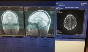
Every MRI scan series begins with a 3-plane localizer scan, also known as a scout scan. The localizer scan acquires a series of images in the axial, sagittal and coronal (X, Y and Z) plane.
After that, the MRI technologist uses the acquired scan sequence to graphically prescribe slice selection for the MRI scan sequences to follow. The 3-plane localizer scan (video below) is a T1-weighted fast gradient echo scan.
Localizer scan sequence scan times average between 12 seconds and 35 seconds to complete.
Four Types of RF pulse sequences utilized For 3-plane Localizer Scan
FGRE Localizer – Fast Gradient Echo Localizer
The FGRE 3-Plane Localizer pulse sequence produces T2-weighted images in the axial, sagittal, and coronal planes. FGRE localizer scans feature higher bandwidth and faster scan times. While the expected tradeoff is lower SNR.
FGRE IR Prep Localizer – Fast Gradient Echo Inversion Recovery
The FGREIR localizer produces quick T1-weighted images in the axial, sagittal and coronal plane. This localizer sequence provides fat suppression and is used when imaging the brain or extremities.
FIESTA Localizer – Fast Inversion Recover Localizer Sequence
The FIESTA localizer pulse sequences produces images of tissue with high t1 and t2 ratios. This localizer scan sequence is ideal when anatomy in scan region has similar inversion recovery times. FIESTA localizer helps avoid blurred edges in the anatomy.
A great example of this would be when imaging requires differentiation between cerebrospinal fluid (CSF), water, and fat. Another example of clinical use would be when imaging requires delineation between muscle, blood vessels and myocardium.
SSFSE MRI Protocol – Short Shot Fast Spin Echo Localizer
Plane Localizer pulse sequence produces T2-weighted images using scan series with customized scan pauses. Single shot fast spin echo scans reduce motion artifact and shorten scan times. No Phase wrap option is unavailable while using SSFSE scan sequences. View the full video here: https://youtu.be/BeV0-b4ZhIo
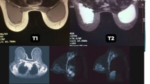
T1 vs T2 MRI Scan Protocol Sounds
Cerebrospinal fluid can cause artifact in brain MRI scans when non-inverted CSF enters into the scan field of view. This can lead to error in diagnosis in the surrounding anatomical structures. To mitigate CSF artifact, the T2 FLAIR sequence provides an inversion slice that is wider than the imaging slice. For more information, see our in-depth T1 vs T2 MRI article.
2. MRI Sounds Produced By Gradient Coil
Gradient sounds are caused by the rapid switching on and off of magnetic fields, while resonance sounds are produced when the RF pulses cause the hydrogen atoms to vibrate. Together, these sounds make up the unique auditory experience of an MRI scan.
The MRI gradient coil produces powerful secondary magnetic fields during the scanning sequence. Below is a list of MRI scans that utilize the MRI gradient coil to produce images.
DWI MRI Scans- Diffusion Weighted Imaging
The DWI-EPI scan sequence is a single shot echo planar imaging pulse sequence. DWI utilizes the gradient coils to provide an echo, as opposed to relying on the RF pulse echo. Additionally, DWI MRI uses partial k-space reconstruction algorithms to reduce scan times. Diffusion Imaging produces images based off of the relative Brownian motion of molecules in the tissue. This scan sequence is ideal when imaging anatomical microstructures. DWI MRI is an ideal scan sequence for brain, breast, prostate, and liver MRI scan techniques.
DWI MRI scans differentiate tissue with restricted diffusion from tissues with normal diffusion coefficients. Diffusion weighted imaging pulse sequences are highly gradient-intensive, T2- weighted scan protocols. DWI MRI creates images based upon measuring the relative Brownian motion of the particles in the tissue.
The water molecules in normal tissue diffuse in a random Brownian motion. This results in a dark or less intense signal in the image. In contrast, the fluid particles in dead tissue experience restricted diffusion, resulting in heightened signal, or lighter images in the tissue. Click here to learn more about DWI MRI scan protocols.
Have you joined the MEDICALIMAGINGSOURCE.COM community? We have a growing library of free articles and videos with the sole mission of providing the most helpful radiology resources.
Subscribe today to stay up to date when we release amazing resources across our platforms.
The video above provides a great example of the sounds made by the gradient coils during an MRI scan.
3. MRI Coldhead Sound
MRI scanners use a cryogenic system to cool the magnets down to extremely low temperatures. This system requires a device called a coldhead that circulates helium gas to cool the magnet. During operation, the coldhead emits a distinct, high-pitched sound that can be heard throughout the scanning room. The frequency of this sound is around 200 Hz, and it is often described as a whistling or buzzing noise. While the sound of the coldhead can be distracting or uncomfortable for some patients, it is an essential part of the MRI system’s operation and is necessary to maintain the magnet superconductive properties.
What Does An MRI Scan Sound Like?
The video below is a compilation of MRI scan sounds from multiple systems. Listen in to various MRI scan protocols to experience what an MRI sounds like.
What is the sound in the background of every MRI scan? The coldhead and compressor units alternate between low pressure and high pressure states. As a result, this produces a rhythmic pumping sound in the background of an MRI scan.
The video below gives a great example of the noise an MRI coldhead assembly produces.
The coldhead compressor functions 24 hours a day to keep the MRI cool. Some MRI scanner configurations shut off the coldhead compressor during the MRI scan sequence. This aids to reduce the likelihood of image artifact.
Conclusion
Magnetic Resonance Imaging scan sounds vary greatly depending on the MRI scanner model and anatomical structure that is being imaged. Additionally, MRI patient protocol sequences are tailored to optimize diagnostic capabilities in each individual patient. MRI scan sounds can change from system to system and patient to patient. It is important to realize the loud MRI noises are provide a safe form of imaging the anatomy. We hope this article was a helpful resource.
Thank you for joining us. Consider subscribing to our blog to stay up to date on the latest resources provided by Medical Imaging Source across all platforms.
Additional Resources
Find in-depth information about MRI artifacts at https://www.medicalimagingsource.com/signa-voyager-mri-artifact-solutions-guide/
MRI (Magnetic Resonance Imaging) Feature Video
Library of informational MRI, CT, and PETCT articles https://www.medicalimagingsource.com/blog
MRI Scan Sounds Playlist on the MRIPETCTSOURCE Youtube channel: https://www.youtube.com/playlist?list=PLtf-HQ_b5QfGNAGSqXmXyNwoXqvcV8zPD
Join the Medical Imaging Source Community!
Subscribe To Our Newsletter To Stay Up To Date With The Latest News, Exclusive Offers, And Giveaways!
The information provided by MRIPETCTSOURCE (“we,” “us,” or “our”) on https://www.medicalimagingsource.com (the “Site”) is for general informational purposes only. All information on the Site is provided in good faith, however we make no representation or warranty of any kind, express or implied, regarding the accuracy, adequacy, validity, reliability, availability, or completeness of any information on the Site. UNDER NO CIRCUMSTANCE SHALL WE HAVE ANY LIABILITY TO YOU FOR ANY LOSS OR DAMAGE OF ANY KIND INCURRED AS A RESULT OF THE USE OF THE SITE OR RELIANCE ON ANY INFORMATION PROVIDED ON THE SITE. YOUR USE OF THE SITE AND YOUR RELIANCE ON ANY INFORMATION ON THE SITE IS SOLELY AT YOUR OWN RISK.