Welcome to our article about MRI top 20 interesting facts! MRI, or Magnetic Resonance Imaging, is a powerful medical imaging technique that has revolutionized the field of diagnostic medicine. It uses a combination of magnetic fields, radio waves, and computer technology to produce highly detailed images of internal body structures.
While many are familiar with MRI fundamentals, there are many fascinating facts about this technology that may surprise you. In this article, we’ll explore 20 interesting facts about MRI that you may not have known before. Whether you’re a healthcare professional, a patient, or someone interested in MRI, you’re sure to learn something new.
Top 20 Interesting MRI Facts
- Top 20 Interesting MRI Facts
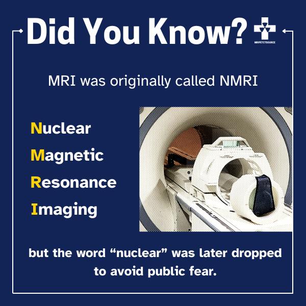
- 1. MRI is Really Nuclear Magnetic Resonance (NMR)
- 2. MRI Uses Non-Ionizing Radiation
- 3. MRI Magnets Are Supercooled to -269 C
- 4. MRI Is Only 50 Years Old.
- 5. MRI Targets Hydrogen Nucleus
- 6. MRI Utilizes NMR To Distinguish Between Different Tissues
- 7. Researchers Use High Field MRI, Up To 11T, Which Come With Greater Risks
- 8. An MRI Is Over 20,000 Times Stronger Than The Magnetic Field
- 9. The Magnetic Field Can Interfere With Medical Implants
- 10. MRI Contrast Is Made Of Paramagnetic Metals
- 11. Functional MRI Shows A Live View
- 12. T1 And T2 MRI Sequences Are Most Commonly Used
- 13. Multicoil Imaging Dramatically Improves Scan Times
- 14. Claustrophobic? Open MRI Is A Great Solution
- 15. MRI Detects Early-Stage Alzheimer’s
- 16. Advanced Algorithms Improve Image Quality With Reduced Scan Times
- 17. AI In MRI Improves Diagnostic Capabilities
- 18. You Can View Your MRI Images The Same Day
- 19. Methods To Reduce The Price Of MRI Scans
- 20. MRI Scan Machines Cost Millions Of Dollars
- Conclusion
1. MRI is Really Nuclear Magnetic Resonance (NMR)
MRI stands for Magnetic Resonance Imaging. It uses the principle of nuclear magnetic resonance (NMR) to produce images of the anatomy. Physicists Felix Bloch and Edward M. Purcell first observed NMR in 1946 independently.
NMR involves nuclei selectively absorbing high frequency radio waves while under an external magnetic field of the same resonance frequency.
2. MRI Uses Non-Ionizing Radiation
Unlike X-rays and CT scans, MRI scans are non-invasive and do not use ionizing radiation. MRI uses strong magnetic fields, radio waves, and computer processing to produce detailed images of internal body structures. It is useful for imaging soft tissues like the brain, muscles, and tendons. These tissues are often not visible on other imaging tests like X-rays, making MRI a valuable tool for this purpose.
3. MRI Magnets Are Supercooled to -269 C
Superconducting coils made of a niobium-titanium alloy generate the magnetic field in an MRI machine. To maintain the superconductive properties, one must cool the coils to temperatures as low as -452 degrees Fahrenheit (4 kelvin) at all times.
4. MRI Is Only 50 Years Old.
The first MRI scan ever performed was a groundbreaking moment in the history of medical imaging. In 1977, Dr. Raymond Damadian and his team at SUNY at Stony Brook used a large, prototype MRI machine to scan a human body for the first time. The entire process took about five hours and only produced a crude black-and-white image of the volunteer’s head.
5. MRI Targets Hydrogen Nucleus
An MRI machine generates radio waves using a high-frequency coil that works like a speaker. These waves excite hydrogen nuclei in the body, causing them to emit a faint signal that the MRI machine detects.
6. MRI Utilizes NMR To Distinguish Between Different Tissues
The signal emitted by the rotating nuclei is used to construct an image of the body. The MRI distinguishes between anatomical structures and produces high-resolution images by detecting different return signals from tissues in the body.
7. Researchers Use High Field MRI, Up To 11T, Which Come With Greater Risks
Developing high-field MRI machines using stronger magnetic fields (7T or higher) has become a topic of interest in recent years. These machines have the potential to provide new insights into the human body’s anatomy and physiology. However, they present engineering challenges, such as increased heating, stronger magnetic fields, and a greater risk of peripheral nerve stimulation.
8. An MRI Is Over 20,000 Times Stronger Than The Magnetic Field
The magnetic field of a 7 tesla magnet is over 140,000 times stronger than the Earth’s magnetic field. At 3T MRI the magnet is over 60,000 times stronger. For comparison, an average scrapyard magnet generates a 1 Tesla magnet and can lift a 3,000 lbs car with ease.
9. The Magnetic Field Can Interfere With Medical Implants
The strong magnetic field in an MRI machine can cause electronic devices, such as pacemakers, to malfunction. That’s why It is important to inform the MRI technologist and imaging staff about any implanted medical devices before undergoing an MRI scan.
Related: MRI and Dental Implants
10. MRI Contrast Is Made Of Paramagnetic Metals
In some cases, a contrast material, such as gadolinium, may be used during an MRI. Contrast agents highlight specific areas of the body to provide higher definition images. MRI contrast is made of materials that have a weak attraction to the MRI magnet. They work by altering the magnetic properties of the surrounding tissues, making them more visible on the MRI image.
11. Functional MRI Shows A Live View
Functional MRI (fMRI) can be used to map brain activity and blood flow. This scan technique uses temporal and spatial resolution to produce images that show neurological changes over time. FMRI is a commonly used diagnostic tool in neuroscience and psychology research. It is used to study brain function as it correlates to behavior.
12. T1 And T2 MRI Sequences Are Most Commonly Used
In MRI, technologists commonly use multi-echo and gradient echo sequences as the two most commonly used imaging techniques. Multi-echo sequences generate T2-weighted images, which detect changes in tissue water content, while gradient echo sequences generate T1-weighted images, which illustrate tissue anatomy.
13. Multicoil Imaging Dramatically Improves Scan Times
Parallel imaging techniques, such as SENSE and GRAPPA, speed up the image acquisition process and reduce overall scan times. These techniques use multiple coils to acquire multiple signals simultaneously, improving the efficiency of the MRI scan.
14. Claustrophobic? Open MRI Is A Great Solution
Recently, open MRI machines have become increasingly popular. They feature a wide open design that provides more space and higher visibility for patients. Open MRI helps to lower incidence of patient anxiety and claustrophobia during the MRI scan.
15. MRI Detects Early-Stage Alzheimer’s
To track the early stages of neurodegenerative diseases like Alzheimer’s and Parkinson’s, researchers are exploring the use of MRI to monitor changes in the brain over time.
16. Advanced Algorithms Improve Image Quality With Reduced Scan Times
Non-standard MRI scan sequences, such as multi-parametric MRI and quantitative MRI, are being developed and used for advanced imaging applications, such as cancer detection and treatment monitoring.
17. AI In MRI Improves Diagnostic Capabilities
AI and machine learning algorithms are developing for use in MRI to improve the accuracy and speed of image analysis and to develop new MRI techniques due to the increasing availability of large datasets.This emerging technology aims to reduce time-consuming tasks of the radiology workflow and maximize accuracy in diagnosis.
18. You Can View Your MRI Images The Same Day
An MRI scan takes varies from 30 minutes to an hour, depending on the type of exam and the area being imaged. While you may not receive your diagnosis the same day, many imaging centers can print your images to a DVD so you have immediate access to your images.
19. Methods To Reduce The Price Of MRI Scans
MRI scans can be expensive. Prices range from a few hundred dollars to several thousand dollars, depending on the type of exam and the location. Patients can net big savings by calling imaging centers in your area to see which is the best imaging option.
20. MRI Scan Machines Cost Millions Of Dollars
Advanced MRI technology comes with a price tag. A 3T MRI scanner costs as much as $3.5 million dollars brand new from the manufacturer. While they may be expensive, these powerful diagnostic imaging systems are revolutionizing our healthcare industry. MRI scan help doctors improve detection of early-stage disease. Additional information available in our MRI Machine Price Guide.
Conclusion
MRI is changing modern medicine by helping doctors diagnose conditions without the need of invasive diagnostic techniques. New MRI technology helps to detect cancers and neurodegenerative disorders at earlier stages than ever before. MRI is a powerful diagnostic tool that will continue to help thousands of patients per year, and only get better with new research.
If you enjoyed this article please make sure to subscribe below to receive a notification when we release new content. We can’t thank you enough for your support!
Related resources
Quick Navigation Links
Join the Medical Imaging Source Community!
Subscribe To Our Newsletter To Stay Up To Date With The Latest News, Exclusive Offers, And Giveaways!
The information provided by MRIPETCTSOURCE (“we,” “us,” or “our”) on https://www.medicalimagingsource.com (the “Site”) is for general informational purposes only. All information on the Site is provided in good faith, however we make no representation or warranty of any kind, express or implied, regarding the accuracy, adequacy, validity, reliability, availability, or completeness of any information on the Site. UNDER NO CIRCUMSTANCE SHALL WE HAVE ANY LIABILITY TO YOU FOR ANY LOSS OR DAMAGE OF ANY KIND INCURRED AS A RESULT OF THE USE OF THE SITE OR RELIANCE ON ANY INFORMATION PROVIDED ON THE SITE. YOUR USE OF THE SITE AND YOUR RELIANCE ON ANY INFORMATION ON THE SITE IS SOLELY AT YOUR OWN RISK.